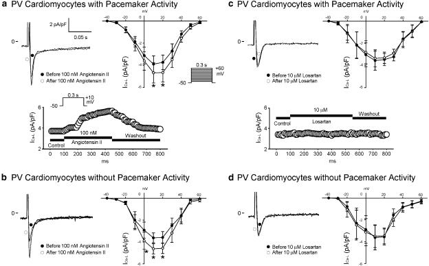
Figure 4.
Effect of angiotensin II and losartan on ICa-L in PV cardiomyocytes. (a and b) The superimposed current traces (depolarization from −50 to +10 mV), time course, and I–V relationship (lower panels) of ICa-L before and after angiotensin II administration in PV cardiomyocytes with (n=10) and without (n=7) pacemaker activity. (c and d) Traces, time course, and I–V relationship before and after the administration of losartan in PV cardiomyocytes with (n=6) and without (n=7) pacemaker activity. *P<0.05 versus before drugs. The insets in the current traces show the various clamp protocols.

