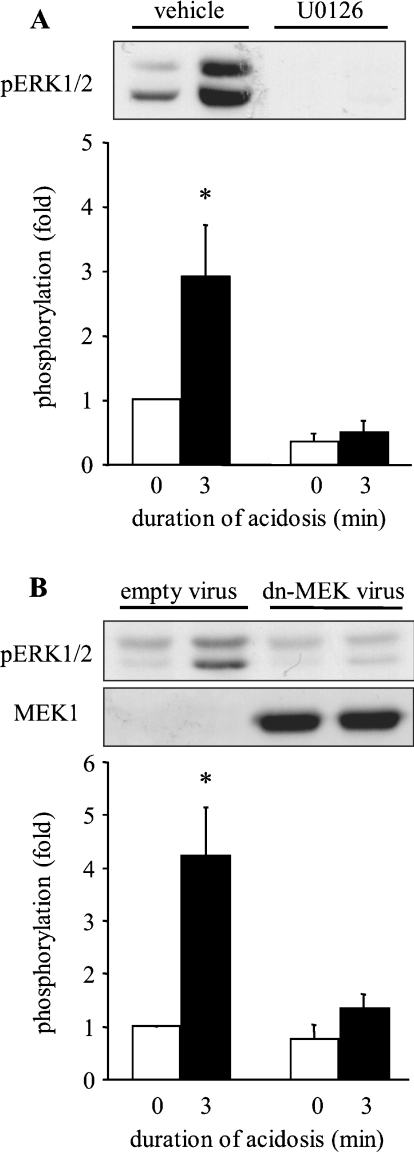
Figure 4. Effect of MEK1/2 inhibition on the activation of ERK1/2 by sustained intracellular acidosis in ARVM.
(A) Western blot of phosphorylated ERK1/2 (pERK1/2) in control cells or following sustained intracellular acidosis in the absence (vehicle) or presence of 3 μM U0126, and quantitative data from four separate experiments. Data are expressed as fold phosphorylation normalized to vehicle control. (B) Western blots of phosphorylated ERK1/2 (pERK1/2) and MEK1 in control cells or following sustained intracellular acidosis 18 h after infection with control adenovirus (empty virus) or D208A-MEK1 (dn-MEK virus), and quantitative data from six separate experiments. Data are expressed as fold phosphorylation normalized to control. *P<0.05 compared with control.

