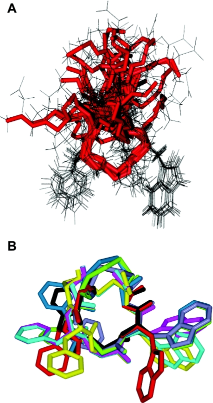
Figure 3. Structure of paionin-1 determined by NMR spectroscopy.
(A) Overlay of the 15 low-energy structures in conformational group number 1, aligned according to the backbone of the CFGWC ring, shown in red. (B) The CFGWC ring of one representative structure from each of the nine conformational groups, each with a distinct colour, aligned according to the backbone.

