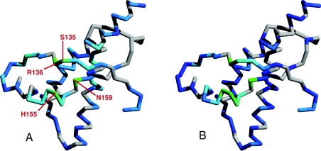
Figure 4. The Cu2+-bound N-terminal domain contacts the C-terminal domain.
The reductions in the heights of backbone [N,HN] signals in the C-terminal domain of PrP, induced by proximity to the Cu2+ bound to the N-terminal domain, are illustrated on the backbone structure by a colour scale, ranging from dark blue to cyan (0–30% peak height reduction) and cyan to green (30–60% reduction). Data is shown for the full-length prion protein loaded with two Cu2+ ions (A) and the truncated PrP91–231 construct, loaded with one Cu2+ ion (B). Residues for which there are no data are coloured grey. Signals from the area of the C-terminal domain that contacts the unfolded N-terminal domain see the largest reductions (coloured green) because they are closer to the Cu2+ ion bound to the unfolded domain. The unfolded domain of the protein and the Cu2+ ion bound to it are omitted for clarity.

