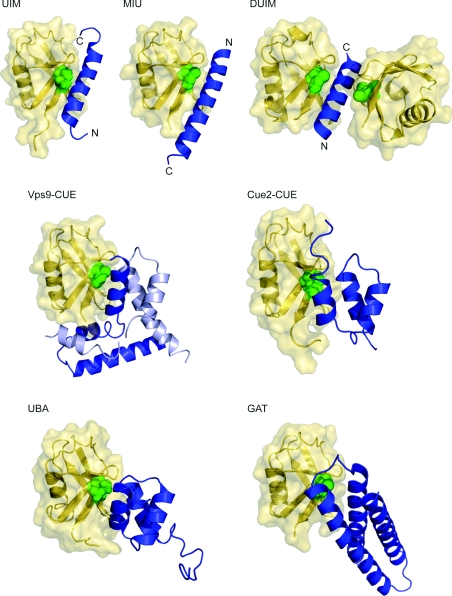
Figure 3. Helical ubiquitin-binding domain structures.
Ubiquitin molecule (yellow) in ribbon and surface representations is shown with corresponding helical domain (blue) in ribbon representation. Ile44, the centre of the hydrophobic recognition patch on the ubiquitin, is shown as green spheres. Ubiquitin molecules are placed in the same orientation as in Figure 1 for comparison. For the UIM, MIU and DUIM structures, both N- and C-termini are marked. Protein data bank identication codes used are as follows: UIM, 1Q0W; MIU, 2FIF; DUIM, 2D3G; Vps9 CUE, 1P3Q; Cue2-CUE, 1OTR; UBA, 1WR1; GAT, 1YD8. Vps9 CUE domain forms a domain-swapped dimer, shown in blue and light blue. The missing part in Vps9 CUE was modelled on the basis of the apo structure.

