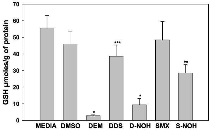
Figure 5.
Glutathione depletion in the presence/absence of parent drugs and their metabolites in NHDF cells: NHDF cells were seeded at a density of 5×10⁵ cells/mL on a 6 well plate and incubated for 3 h with 1.5 mM of parent drug or metabolite. Media with NHDF cells alone were used to measure the basal GSH content, DMSO (0.25%) was used as the control, and DEM 0.4 mM was used as a positive control for GSH reduction. After 3 h incubation, the cells were washed with ice cold PBS and treated with 1 ml ice cold lysis buffer. Cells were lysed, extracted and analyzed for GSH content as described in Materials and Methods. GSH contents were determined using a standard curve generated from known concentrations and expressed as μmoles/g of protein. Data presented as mean (+SD) of four separate incubations for each condition. Results were analyzed using ANOVA with Holm-Sidak method for multiple pairwise comparisons. *p<0.05 compared to media, DMSO, DDS, SMX and SNOH; **p<0.05 compared to media, DMSO, DDS, and SMX; ***p<0.05 compared to media, DMSO, and SMX.

