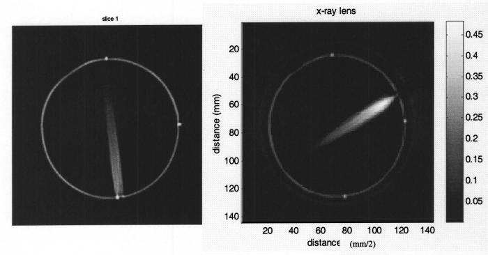
FIG. 12.
Optical-CT images of reconstructed attenuation coefficients in 120 kVp irradiations. In the left image the unfocused x-ray beam is collimated to 5 mm diameter utilizing a lead sheet. In the right image, the x-rays have been focused to a point in the gel dosimeter utilizing the x-ray lens in Fig. 4.

