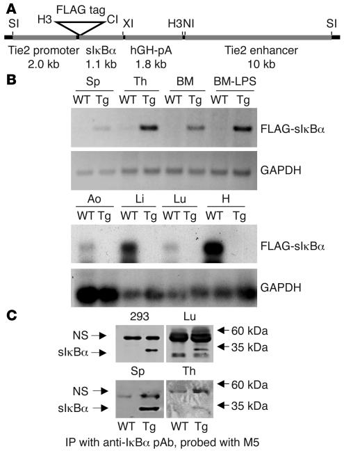
Figure 1. Generation of transgenic mice.
(A) The transgenic construct consisted of the Tie2 promoter, dominant negative FLAG-tagged sIkBαS32A/S36A, hGH polyadenylation cassette (hGH-pA), and the Tie2 enhancer. Restriction sites for SalI (SI), HindIII (H3), ClaI (CI), XbaI (XI), and NcoI (NI) are shown. (B) RT-PCR analysis for sIκBα expression in RNA from transgenic (line 2) and WT heart (H), lung (Lu), liver (Li), aorta (Ao), spleen (Sp), thymus (Th), and BM. The PCR product was visualized either by UV fluorescence (upper 2 panels) or hybridization with IκBα or GAPDH [32P]dCTP-radiolabeled probes (lower 2 panels). One set of BM was harvested from mice 7 days after LPS stimulation (2 μg/g). (C) IκBα and sIκBα protein expression was evaluated by a sensitive immunoblotting (anti-FLAG M5 mAb; Sigma-Aldrich)/immunoprecipitation (IκBα polyclonal Ab [pAb]; Santa Cruz Biotechnology Inc.) assay in lung, splenic, and thymic extracts prepared from transgenic mice (line 4). Extracts prepared from HEK 293T cells transiently transfected with the sIκBα expression vector served as a positive control. A nonspecific (NS) band (recognized by M5 mAb) and size standards are indicated.

