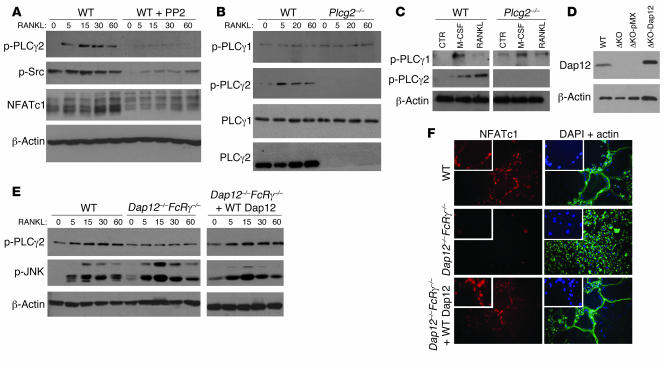
Figure 6. PLCγ2 is activated by RANKL via Dap12/FcRγ in an SFK-dependent manner.
(A) WT OC precursors (preOCs; BMMs grown in RANKL-containing media for 2 days) cultured with the SFK inhibitor PP2 (5 μM) or vehicle (DMSO) were stimulated with RANKL and subjected to Western blot analysis to detect phosphorylated levels of PLCγ2, Src, and NFATc1. β-Actin served as control. (B) PLCγ1 and PLCγ2 phosphorylation in response to RANKL were measured by Western blot analysis in WT and Plcg2–/– preOCs. PLCγ1 and PLCγ2 levels are shown. (C) PLCγ1 and PLCγ2 phosphorylation in response to 5 minutes of treatment with either M-CSF or RANKL in WT and Plcg2–/– preOCs. β-Actin served as control. (D) Expression levels of endogenous Dap12 and Flag-tagged Dap12 retrovirally transduced in Dap12–/–FcRγ–/– BMMs are shown. ΔKO, Dap12–/–FcRγ–/–. (E) PLCγ2 phosphorylation was measured by Western blot analysis in WT, Dap12–/–FcRγ–/–, or Dap12–/–FcRγ–/– preOCs reconstituted with WT Dap12 stimulated with RANKL for the indicated times. Phospho-JNK is also shown. β-Actin served as loading control. (F) Nuclear localization of NFATc1 in WT and Dap12–/–FcRγ–/– OCs retrovirally transduced with pMX or Flag-tagged Dap12 is shown in red (left panels). Actin staining is shown in green, and nuclei, stained with DAPI, are shown in blue (right panels) (objective, ×20). Enlarged images (2.5-fold) show nuclear localization of NFATc1 (red) and nuclei stained with DAPI (blue) of representative cells located in the center of the photographed field.

