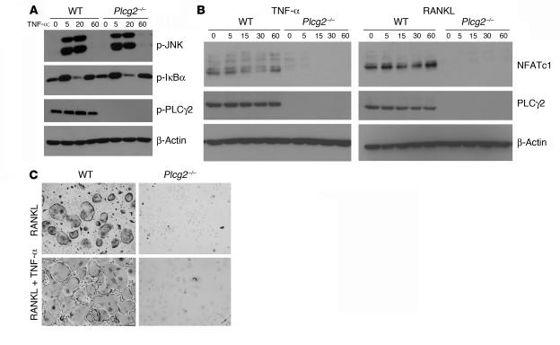
Figure 8. TNF-α cannot correct the Plcg2–/– OC defect.
(A) WT and Plcg2–/– BMMs were stimulated with TNF-α (10 ng/ml) with time. Phosphorylated JNK, IκBα, and PLCγ2 were examined. β-Actin served as control. (B) WT and Plcg2–/– BMMs were cultured for 3 days with RANKL (100 ng/ml) and M-CSF (10 ng/ml). On day 3 TNF-α was also added to the culture media. Cells were fixed and TRAP stained at day 7. Magnification, ×200. (C) WT and Plcg2–/– preOCs were stimulated with TNF-α or RANKL for the indicated times, and expression levels of NFATc1, PLCγ2, and β-actin were determined.

