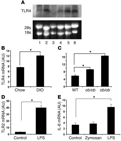
Figure 3. Adipocytes express functional TLR4, and TLR4 expression is increased in adipose tissue of obese models.
(A) Adipocytes express TLR4. Northern blotting was used to detect TLR4 mRNA expression. Lane 1: 3T3-L1 preadipocytes; lane 2: 3T3-L1 adipocytes; lane 3: stromal-vascular cells; lane 4: mouse adipocytes; lane 5: mouse adipose tissue; lane 6: RAW264.7 macrophages. (B and C) TLR4 mRNA expression is increased in fat pads from DIO and ob/ob and db/db mice (n = 4; *P < 0.05). TLR4 mRNA was measured using real-time RT-PCR. (D) The TLR4 agonist LPS induces TLR2 expression in adipocytes. (E) LPS but not zymosan stimulates IL-6 mRNA expression in 3T3-L1 adipocytes. 3T3-L1 adipocytes were treated with 100 ng/ml LPS or 40 μg/ml zymosan for 8 hours. Real-time RT-PCR was conducted to measure TLR2 and IL-6 mRNA levels. Data are expressed as mean ± SEM; n = 6, *P < 0.05.

