Figure 5.
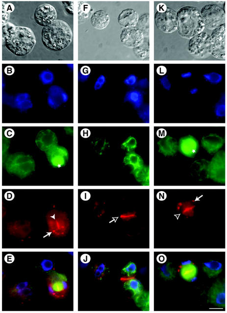
NPSN11 is localized at the cell plate during cytokinesis. Arabidopsis suspension-cultured cell protoplasts (A, F, and K) were double immunolabeled with antibodies directed against α-tubulin (α-tub) to visualize phragmoplast (marked by stars), cortical microtubules (C, H, and M), and either affinity-purified NPSN11 (D and I) or KNOLLE antisera (N). Nuclei in dividing and non-dividing cells were revealed by staining with 4′,6′-diamidino-2-phenylindole (DAPI; B, G, and L). Electronically merged images of B through D, G through I, and L through N are shown in E, J, and O, respectively. NSPN11 positive cell plate (solid arrowhead) and new plasma membrane (empty arrow) are visible in D and I, respectively. N shows the localization of KNOLLE at the cell plate (empty arrowhead). Arrows in D and N indicate NSPN11- and KNOLLE-containing intracellular organelles. Scale bar in A through O = 10 μm.

