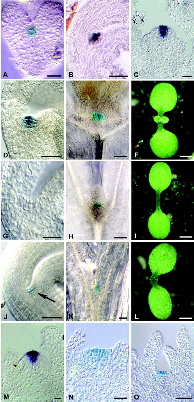
Figure 1.
Dependence of CLV3 expression on WUS and STM. A, GUS-stained heart stage CLV3::GUS embryo. B, CLV3 expression in a bent cotyledon stage embryo, detected by in situ hybridization with a CLV3 probe. C, CLV3 expression in a CLV3::GUS inflorescence meristem, detected by in situ hybridization with a GUS probe. D, GUS-stained mature CLV3::GUS embryo; compare with B. E, GUS-stained wild-type seedling 10 d after germination (d.a.g.). F, Wild-type seedling 10 d.a.g. The first leaf pair is visible. G, GUS-stained mature CLV3::GUS/wus-1 embryo. CLV3 expression is not detectable. H, GUS-stained CLV3::GUS/wus-1 seedling 10 d.a.g. I, wus-1 seedling 10 d.a.g. J, GUS-stained mature CLV3::GUS/stm-11 embryo showing CLV3 expression (arrow). K, GUS-stained CLV3::GUS/stm-11 seedling 10 d.a.g. showing CLV3 expression. L, An stm-11 seedling 10 d.a.g. has formed cotyledons, but no SAM is visible. M, Wild-type CLV3::GUS axillary meristem 21 d.a.g. GUS RNA is detected by in situ hybridization. N, GUS-stained CLV3::GUS/wus-1 axillary meristem 21 d.a.g. O, GUS-stained CLV3::GUS/stm-11 axillary meristem 21 d.a.g. Scale bars in F, I, and L = 1 mm; in all other figures, scale bars = 20 μm.

