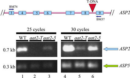
Figure 2.
Semiquantitative RT-PCR of ASP2 mRNA in aat2 mutants. A schematic drawing of the ASP2 gene including exons 3 through 9 (blue) and introns (pink) indicates where the T-DNA insertion is located (red) and the oligonucleotide primers on the ASP2 gene used in the RT-PCR (BM74 and BM37). PCR was performed for 25 cycles (lanes 1–3) or for 30 cycles (lanes 4–6) on wild type (lanes 1 and 4), aat2-T (lanes 2 and 5), and aat2-5 (lanes 3 and 6). Co-Amplification of ASP5 was used as a positive control in the same PCR reaction tube.

