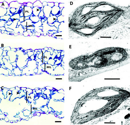
Figure 5.
Histological analysis of partially rescued rsy3 leaves. Separate sections of leaves shown in Figure 4E were taken for histological analysis (A–C) and for transmission electron microscopic (TEM) analysis of chloroplasts (D–F). Tissue section (A) and TEM of chloroplast (D) from morphologically wild-type green plants (RSY3/RSY3;tE989). Leaf tissue section (B) and TEM of a chloroplast (E) from a pale-yellow region of partially rescued rsy3 mutant plants (rsy3/rsy3; tE989/tE989). Leaf tissue section (C) and TEM of a chloroplast (F) from a green region of partially rescued rsy3 mutant plants (rsy3/rsy3; tE989/tE989). Arrowheads point to chloroplasts. PL, Palisade mesophyll layer; ML, spongy mesophyll layer; g, grana. Bars = 100 μm in A through C and = 0.15 μm in D through F.

