Fig. 2.
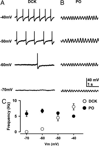
Repetitive firing of DCK neurons depends on the membrane potential. (A) Representative recordings with a high-potassium intracellular solution in the current clamp configuration showing the discharges of a DCK neuron at membrane potentials of −70, −60, −50, and −40 mV. Note that the firing frequency increased with depolarization. Each individual action potential was followed by a large, long-lasting afterhyperpolarization. Action potentials in this figure were truncated to illustrate the afterhyperpolarization amplitude and time course. (B) Representative patch recordings from a PO neuron showing subthreshold oscillations at the same membrane potentials as in A. Note that the oscillation frequency (5 Hz) hardly changed with membrane potential, although the amplitude changed. The DCK neuron in A and the PO neuron in B were recorded in the same slice. (C) Mean firing frequency (±SEM) of DCK neurons (n = 11) and mean frequency (±SEM) of subthreshold oscillations of PO neurons (n = 10) as a function of membrane potential. The membrane potentials are the steady-state values reached after injection of a slowly depolarizing ramp current. Note that the frequency of DCK neurons (open circles) increased linearly with membrane potential whereas the frequency of PO subthreshold oscillations (filled circles) remained near 5–7 Hz.

