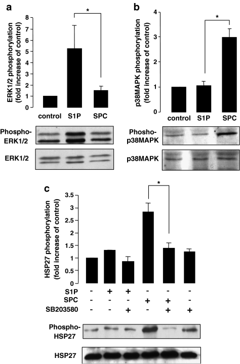
Figure 1.
S1P- and SPC-induced phosphorylation of MAP kinases and HSP27 in rat cerebral arteries. (a) Representative immunoblots showing changes in phosphorylation of ERK1/2 by S1P and SPC in rat cerebral arteries. The bar graph represents fold increase in phosphorylation of ERK1 and ERK2 combined compared to basal levels. S1P (5 μM) significantly increased phosphorylation after 15 min stimulation (n=6). SPC (10 μM), however, did not significantly increase ERK1/2 phosphorylation after 15 min stimulation. (b) Representative immunoblots and bar graph demonstrating increased phosphorylation of p38MAPK by S1P and SPC in rat cerebral arteries. After 15 min of stimulation, SPC (10 μM) significantly increased phosphorylation (n=6) while S1P (5 μM) did not significantly increase p38MAPK phosphorylation (n=6). (c) Representative immunoblots and bar graph showing the fold increases in phosphorylation of HSP27 by S1P and SPC in the rat cerebral artery. Following 15 min stimulation, SPC (10 μM) significantly increased HSP27 phosphorylation; however, S1P (5 μM) did not increase HSP27 phosphorylation (n=5). Preincubation with the p38MAPK inhibitor, SB203580 (30 μM), for 30 min, the SPC (10 μM)-induced phosphorylation was significantly inhibited (n=5). Data shown are mean±s.e.m. Asterisk denotes P<0.05.

