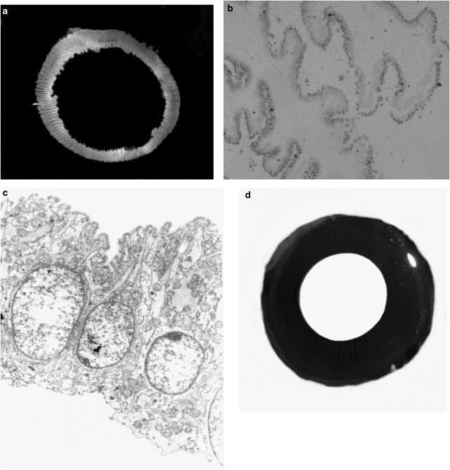
Figure 1.
(a) Photograph of the intact ring of NPE cells isolated from the porcine eye. (b) Histological section of the isolated NPE ring showing minimal contamination by PE cells. Examination under light microscope shows that the tissue samples comprised more than 99% NPE cells. (c) Electron microscopic section (× 4350) shows a single layer of cells with no apparent PE cells. Note that the basolateral side of the NPE is uppermost. There seems to be no obvious contamination with PE cell remnants or pigment granules. (d) Cross-section of the porcine eye exposing the ciliary body from behind. The ciliary body appears as radial black ridges that extend onto the posterior surface of the iris. The pupil appears as the disc in the center of the image. Abundant pigmentation is clearly visible in the intact ciliary body.

