
Figure 5.
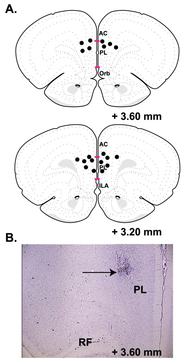
Sites of intra-mPFC anisomycin infusions. A, Depiction of the mPFC on atlas plates at two different rostral-caudal planes modified from Swanson (2004). Non-redundant sites of infusion termini (marked by black circles) in the mPFC from a representative sample of animals. The horizontal bars mark the borders of the prelimbic (PL) cortex. None of the infusion tracks terminated in the infralimbic (IL) cortex. B, A representative photomicrograph of a coronal section stained with cresyl violet indicating the terminus of an infusion needle (shown by arrow). AC, anterior cingulate; ILA, infralimbic cortex; PL, prelimbic cortex; RF, rhinal fissure; Orb, medial orbital cortex.