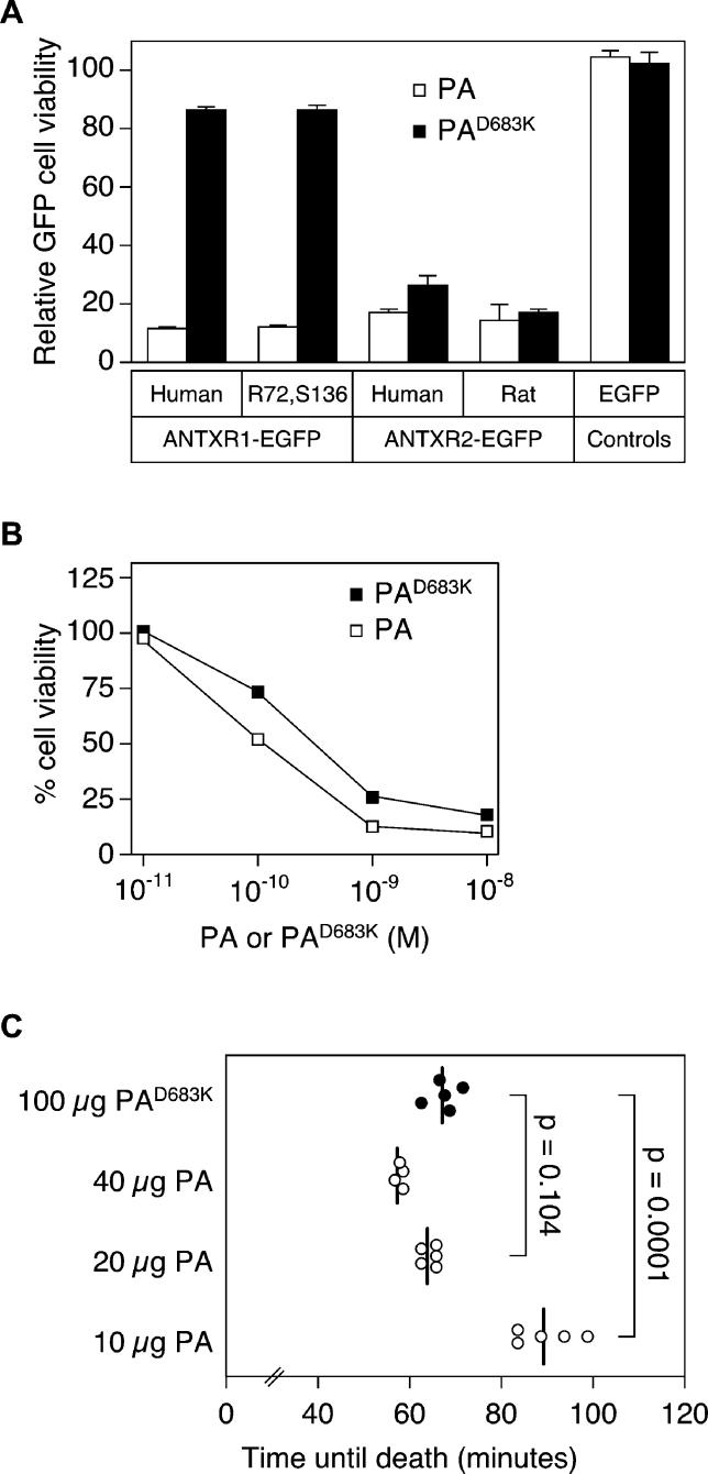
Figure 4. PAD683K Supports Lethal Toxin-Mediated Killing of Rats via rANTXR2.
(A) Triplicate samples of CHO-R1.1 cells transiently expressing the different receptor-EGFP proteins were incubated with 10−10 M LFN-DTA and with either 10−8 M wild-type PA or 10−7 M PAD683K or without PA. These amounts of wild-type and mutant PA proteins were used because they gave rise to maximal levels of killing when incubated with cells expressing human ANTXR2 (Figure 1D). Cell viability was measured by counting the percentage of live EGFP-positive cells in the sample by flow cytometry and is expressed as described in the Figure 3A legend.
(B) Triplicate samples of CHO-R1.1 cells transiently expressing ANTXR1R72,S136-EGFP were incubated with toxin and analyzed as in (A) except that increasing amounts of either the wild-type or mutant PA protein were used.
(C) Groups of five rats each were injected by jugular vein cannula with 8 μg of LF and either 100 μg of PAD683K or 10 to 40 μg of PA and time until death postinjection was recorded. One rat in the 40 μg PA group died immediately after injection from pulmonary embolism and was excluded from analysis. Each data point represents a single animal, and the vertical line represents the mean for each group. P-values were determined by Student's unpaired t-test. Control rats injected with PBS all survived (unpublished data).

