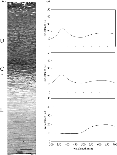
Figure 1.
(a) Toluidine blue stained histological transverse section of beak horn, cut parallel to the short axis, with the outer surface at the top, showing distinct upper (U), central (C) and lower (L) superposed regions. Scale bar, 25 μm. (b) Reflectance spectra obtained on intact and scraped beak horn. The upper spectrum was obtained on intact beak horn; the middle spectrum was obtained on scraped beak horn after partial removal of the upper region; the lower spectrum was obtained when the entire upper region had been removed by scraping, leaving the central and lower regions intact.

