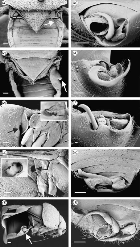
Figure 2.
Coridromius female paragenitalia (a,c,e,g,i) and corresponding male genitalia (b,d,f,h,j). Paragenital structures are indicated by arrows. C. chinensis (a,b): in the female, the first right lateral tergite is slightly swollen and thinly sclerotized (a). The site of copulation (not visible) is high on the anterior margin of the first visible ventral sternite, into which the male's scythe-like paramere (b) is inserted. Undescribed species from the Philippines (c,d): the paragenitalia of this species include swelling of both the dorsolateral tergites and the corresponding abdominal sternites. Undescribed species from Tahiti (e,f): in this species the female ectospermalege takes the form of a prominent copulatory tube (e), which opens into a thinly sclerotized cavity (e inset). C. zetteli (g,h): in C. zetteli, males have a corkscrewed intromittent organ (h) which is inserted into the female copulatory tube (g) located on the right side of the abdomen. This corkscrewed copulatory tube is punctured at its apex (g inset), indicating the point of insemination. Undescribed species from South Africa (i,j): in this species both females and males possess unusual ‘cup and bulb’ paragenitalia (i), the function of which is unknown. All scale bars, 100 μm.

