
FIG. 2.
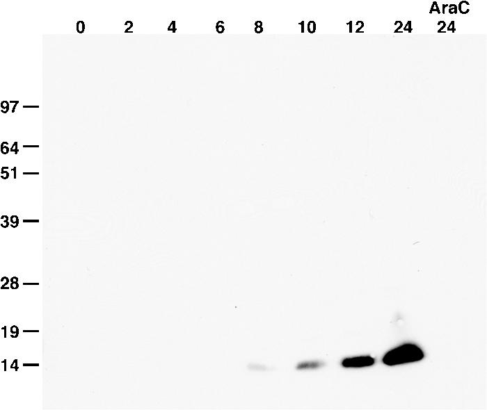
Kinetics of F9 expression. BS-C-1 cells were infected with VACV for the hours indicated at the top of the figure, harvested in the presence of N-ethylmaleimide, and analyzed by nonreducing SDS-PAGE and Western blotting with a rabbit polyclonal antibody specific for F9 followed by horseradish peroxidase-conjugated anti-rabbit antibody. One infection was performed in the presence of AraC, and cells were harvested at 24 h. Bands were detected by chemiluminescence. Numbers at left show molecular masses in kilodaltons.