
FIG. 6.
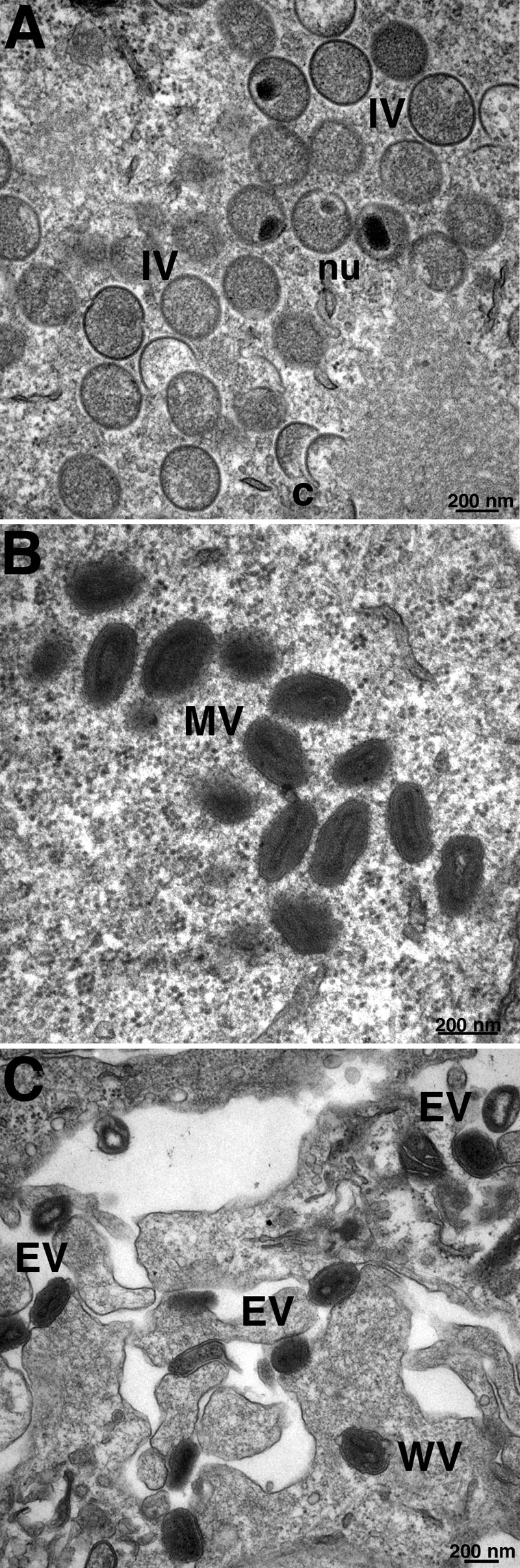
Electron microscopy of cells infected with vF9Li in the absence of IPTG. BS-C-1 cells were infected with vF9Li at a multiplicity of 4 PFU per cell in the absence of IPTG. After 20 h, the cells were processed for transmission electron microscopy as previously described (41). (A) Crescents (c) and IVs with visible nucleoids (nu). (B) MVs. (C) Wrapped virions (WV) and EVs. Bars, 200 nm.