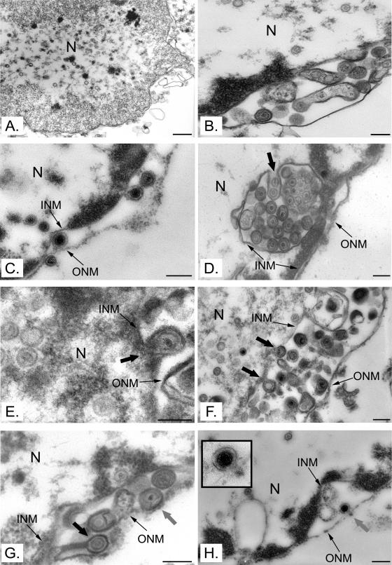
FIG. 8.
Viral egress in vitro. Nuclei isolated from HeLa cells infected with HSV-1 17+ were analyzed by Epon embedding and electron microscopy. Capsids were present in the nuclei (A) and the perinuclear space (B and C). Capsids seemingly in the process of acquiring an envelope at the inner nuclear membrane (large black arrows in panels D to G) or interacting with the outer nuclear membrane (large gray arrows in panels G to H) were observed. Bars represent 500 nm in panel A and 200 nm in all other panels. N, nucleus; INM, inner nuclear membrane; ONM, outer nuclear membrane.

