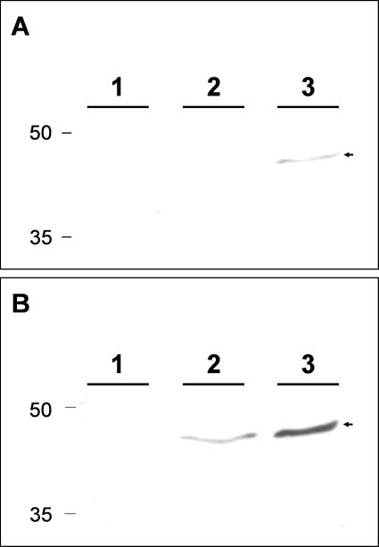
FIG. 10.
Detection of HOXA9 protein in MeWo cells. MeWo cell monolayers were passaged and incubated for 3 days. At time zero, one monolayer was harvested (lane 1). Two other monolayers were incubated for an additional 48 h before harvesting; one of these was untreated (lane 2), and one was treated with HMBA (lane 3). Cell lysates were separated on a 12% acrylamide gel and transferred to a polyvinylidene difluoride membrane. All three monolayers were subsequently probed for the HOXA9 protein with a murine MAb. The membrane was exposed for 4 min (A) or 20 min (B). The HOXA9 protein is designated with an arrowhead.

