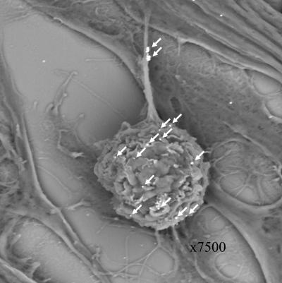
FIG. 4.
Scanning electron micrograph of an inoculum cell. At 24 hpi, an infected monolayer was examined by scanning electron microscopy. The sample had been treated with a murine anti-gC MAb, followed by an immunogold-labeled anti-mouse antibody. After silver enhancement, 12 gC-positive beads were clearly seen on the surface of the inoculum cell, and two clusters of beads were observed on an extension (arrows). Compare this with the gC immunostaining in Fig. 3.

