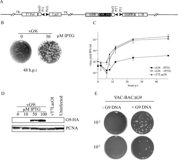
FIG. 2.
Correlation of G9 expression with virus replication. (A) Construction and characterization of a G9-inducible virus. The diagram shows relevant segments of the recombinant vG9i genome. T7 pol, bacteriophage T7 RNA polymerase; PT7, bacteriophage T7 promoter; lacO, E.coli lac operator; lacI, E.coli lac repressor; P11, vaccinia late promoter; Pe/l, P 7.5 early/late promoter; TK, thymidine kinase locus. (B) Plaque formation in the presence and absence of inducer. BS-C-1 monolayers were infected with the vG9i in the presence of 0 or 50 μM IPTG. At 48 h postinfection (h.p.i), the cells were stained with crystal violet. (C) Single-step virus yield in the presence and absence of inducer. vG9i or the parent virus vT7LacoOI was adsorbed to BS-C-1 cells (5 PFU/cell) in the presence or absence of IPTG for 60 min at 37°C. After 1 h, the residual inoculum was removed and the cells were washed and overlaid with medium with or without IPTG. Cells were then harvested at zero time or at the indicated times postinfection (p.i.). The crude lysates were treated with 125 μg/ml of trypsin for 30 min at 37°C to disperse virus particles. Virus yields were determined by plaque assay with 50 μM IPTG. Averages of data for two experiments with error bars are shown. (D) Western blot. BS-C-1 cells were infected with 5 PFU of vG9i in the presence of the indicated amounts of IPTG. After 24 h, whole-cell extracts were analyzed by Western blotting with an anti-HA MAb to detect G9 and anti-proliferating cell nuclear antigen (PCNA) rabbit antibody as a loading control. (E) Attempt to delete the G9R ORF. CV-1 cells were infected with FPV and transfected with the VAC-BACΔG9 plasmid. After 7 days, the cells were lysed and the presence or absence of infectious virus was determined by plaque assay. In parallel, another plate of CV-1 cells was infected with FPV and transfected with the VAC-BACΔG9 plasmid plus or minus DNA containing the G9 wild-type sequence and flanking region and assayed in the same manner.

