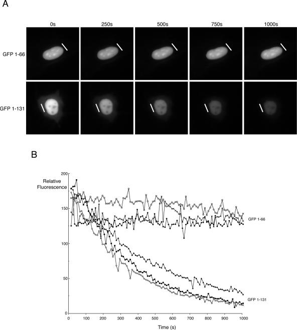
FIG. 7.
Analysis of nucleocytoplasmic shuttling by FLIP. (A) HEp-2 cells were transfected with GFP fused to either residues 1-66 or residues 1 to 131 of bUL47. Twenty hours later, the expressing cells were examined using a Deltavision RT imaging system, and individual cells were chosen for FLIP analysis. The laser module was then used to carry out sequential photobleaching events of the area denoted by the white line. This area was exposed to the laser every 10 s for 100 repeats. An image of the field was acquired after each bleach event to determine the loss of fluorescence in the nucleus of the cell. (B) For each fusion protein, the relative fluorescence in the nuclei of three individual cells that had been subjected to FLIP was quantitated using NIH Image software and plotted against time.

