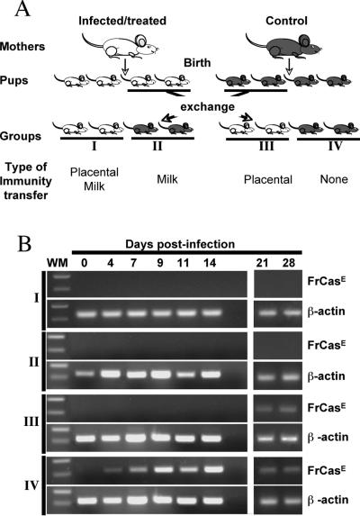
FIG. 2.
Experimental scheme and analysis of proviral DNA in splenocytes. (A) Experimental scheme. Two infected/treated and two control noninfected/nontreated mice were mated at the same time to ensure synchronized deliveries. Immediately after birth and before breastfeeding could occur, the pups were distributed as follows: eight neonates from each litter were kept with their natural mothers for nursing, whereas another eight were transferred to a foster mother in order to have animals born to infected/treated mice nursed by control mice and vice versa. All neonates were infected with FrCasE 3 days after birth and followed for signs of illness, anti-FrCasE antibodies, and viral propagation using several criteria (see the text). Analysis at early time points required the sacrifice of two animals per time point. (B) RT-PCR detection of FrCasE Env mRNA in the spleen. Two animals per group were sacrificed at the indicated times. The spleens were recovered and pooled for RNA preparation. Subgenomic FrCasE Env mRNA accumulation was assayed by RT-PCR as described in Materials and Methods. β-Actin was used as an internal invariant amplification standard. WM, molecular weight marker.

