FIG. 8.
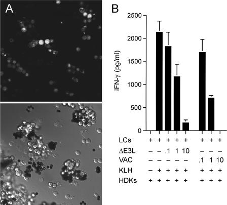
Abortive vaccinia virus infection of primary LCs inhibits their antigen presentation function. (A) LCs were purified from female BALB/c mouse epidermis with anti-I-Ad antibody, followed by incubation with magnetic beads coated with goat anti-mouse immunoglobulin G antibody. They were infected with vaccinia virus NP-S-EGFP at a multiplicity of 10. At 24 h postinfection, the cells were imaged with a Zeiss laser scanning confocal microscope. The bright-field image is shown in the bottom half of the panel. Green fluorescence of the same field is shown in the top half of the panel. (B) Antigen presentation. Purified primary LCs were infected with vaccinia virus WR or ΔE3L at a multiplicity of 0.1, 1, or 10 or mock infected. Cells bound to magnetic beads were washed to remove unadsorbed virus, incubated overnight with the KLH antigen, and then washed to remove KLH. LCs (1 × 10⁴) were then coincubated with HDK-1 cells (5 × 10⁴) for 72 h. The IFN-γ concentration in the medium was measured by ELISA. Each datum represents the average of three antigen presentation assays with standard deviations shown.

