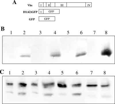
FIG. 6.
Membrane association of D1(42)GFP in the presence and absence of infection. (A) Schematic representation of the D1(42)GFP construct used to analyze domain 1 of Vhs. (B and C) Membrane association, in duplicate, of (B) GFP or (C) D1(42)GFP in the presence (lanes 5 to 8) and absence (lanes 1 to 4) of infection. Lanes 1, 3, 5, and 7 represent the membrane fraction, whereas the corresponding rest of the gradient is in lanes 2, 4, 6, and 8.

