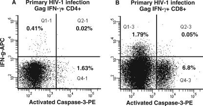
FIG. 2.
Lack of apoptosis of Gag-specific CD4+ T cells in vitro during PHI. Following stimulation with Gag peptides in the intracellular cytokine assay, CD4+ (A) and CD8+ (B) T cells were simultaneously stained with monoclonal antibodies to IFN-γ and activated caspase-3. Representative histograms for one subject out of four consecutive subjects with PHI are shown. Also shown are percentages of cells in quadrants.

