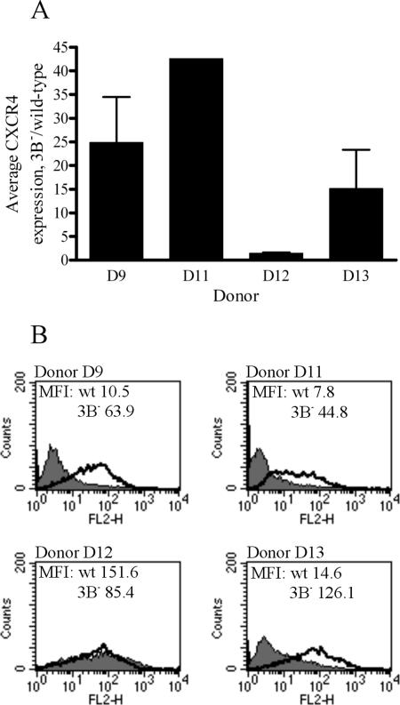
FIG. 9.
RNA and cell surface protein expression of CXCR4 in 3B− LCLs from multiple donors. (A) Average expression levels of CXCR4 mRNA in 3B− LCLs, relative to expression levels in wild-type LCLs. Multiple independent wild-type and 3B− LCLs were generated from four different donors, and quantitative RT-PCR for CXCR4 was performed on RNA isolated from these cells. Average CT values for CXCR4 were normalized to GAPDH levels, and data displayed are changes (n-fold) in expression levels in 3B− LCLs relative to expression levels in wild-type LCLs. Data are means ± standard deviations for independent experiments performed in triplicate on independently derived 3B− LCLs. (B) Cell surface expression of CXCR4 protein in wild-type (wt) and 3B− LCLs from different donors. Wild-type and 3B− LCLs were stained for CXCR4 expression by using a PE-conjugated antibody directed against CXCR4 (clone 1D9). A PE-conjugated, isotype-matched antibody was used as a control. The shaded area shows CXCR4 staining of wild-type LCLs, and the bold line represents staining of 3B− LCLs. The mean fluorescence intensities (MFI) of each sample are indicated on their respective graphs.

