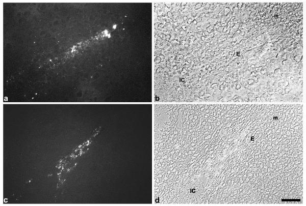
Figure 1.
Immunostaining for TNFalpha. Immunoreactivity to TNFalpha is strongly expressed in the luminal epithelium close to the implantation chamber on day 5.5 pc (a, b). Staining for TNFalpha is enhanced on day 6.5 pc in the luminal epithelium (c, d). IC, implantation chamber; E, luminal luterine epithelium; m, mesometrial side. Bar represents 50 μm in a-d.

