
Figure 3.
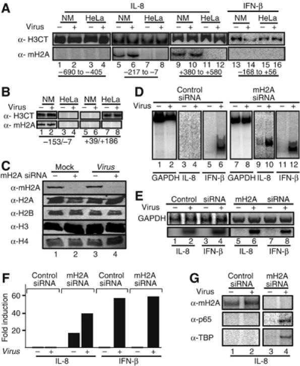
The histone variant macroH2A marks the IL-8 gene for repression in B cells. (A) HeLa and Namalwa cells were mock or virus infected for 6 h followed by formaldehyde crosslinking and immunoprecipitation using antibodies specific for macroH2A1.2 and the carboxyl-terminus of histone H3. The precipitated chromatin-bound DNA was detected using the indicated set of primers amplifying the upstream IL-8 region (−690 to −405, lanes 1–4), the enhancer/promoter (−217 to −7, lanes 5–8) and the coding region (+380 to +580, lanes 9–12) or the IFN-β enhancer (lanes 13–16). The relative position of the amplified regions is shown at the bottom of the gel. (B) Same as in (A) except the chromatin used was composed of mononucleosomes prepared after digestion with micrococcal nuclease. (C) A Western blot using whole-cell extracts prepared from mock- or virus-infected HeLa cells transfected with a control siRNA (lanes 1, 3) or the macroH2A-specific siRNA (lanes 2 and 4). Our transfection efficiency in HeLa cells is approximately 90%. The abundance of macroH2A1.2 and histones H2A, H2B, H3 and H4 was determined by Western blotting using specific antibodies. (D) Namalwa cells were transfected with the control (lanes 1–6) or the macroH2A-specific siRNA (lanes 7–12) and 48 h later they were infected with Sendai virus for 6 h. The isolated RNA was used as a template in RT–PCR reactions using primers detecting GAPDH, IL-8 and the IFN-β-coding regions. (E) Same as in (D) except the RNAs used in the RT–PCR reactions were prepared from HeLa cells transfected with the indicated siRNAs. (F) Namalwa cells were transfected with luciferase-based reporter plasmids bearing the IL-8 or the IFN-β enhancer promoter along with control or macroH2A siRNAs. At 48 h following transfection, the cells were mock or virus infected for 10 h and the luciferase activity was determined. The average of three experiments is shown and the variability from experiment to experiment was less than 20%. (G) Namalwa cells were transfected with the IL-8 reporter as in (F). At 6 h after virus infection, the cells crosslinked with formaldehyde and the isolated chromatin was immunoprecipitated using the indicated antibodies. PCR was used to detect the precipitated IL-8 promoter fragments using plasmid-specific primers.