
Figure 1.
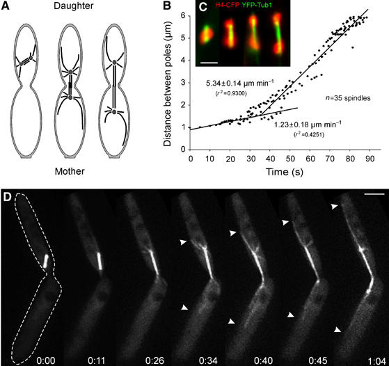
Spindle elongation in anaphase in U. maydis. (A) Overview of late mitotic stages in U. maydis. Anaphase is initiated in the daughter cell, and spindle elongation segregates the chromosomes into the daughter and mother cells (modified from Steinberg et al, 2001). (B) Linear regression analysis of spindle elongation during anaphase. Spindle elongation starts with a slow phase, but rapidly increases when spindles reach a length of 1.5–2 μm. The graph is based on the analysis of 35 spindles that were aligned; each spindle was observed for 30 s. (C) DNA separation in strain FB1H4C_YT, which expresses a CFP-H4 fusion protein (red) and YFP-α-tubulin (green). Initial steps of chromosomes separation are categorized and stages 1–4 correspond to Figure 5C. Note that images were taken from different spindles. Bar: 2 μm. (D) Time-lapse fluorescence microscopy of spindle elongation and spindle positioning in U. maydis strain FB2GT using GFP-α-tubulin-labeled MTs. Note that rapid spindle elongation coincides with the appearance of long astral MTs at both spindle poles (arrowheads) that slide along the cortex while the spindle elongates. The edge of the cell is indicated at time 0 by a dotted line. Elapsed time in minutes and seconds is indicated. Bar: 3 μm.