
Figure 1.
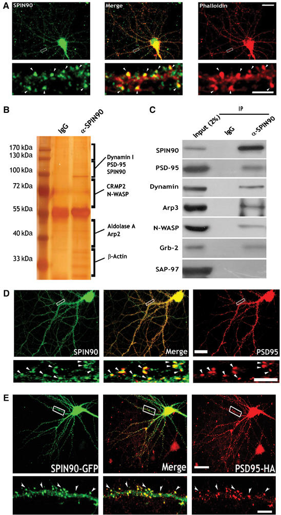
SPIN90/WISH is expressed in dendritic spines and colocalizes with actin and PSD-95. (A) Colocalization of SPIN90/WISH and actin in cultured hippocampal neurons. Neurons at 17DIV were stained with anti-SPIN90/WISH antibody (green) and phalloidin (red). A high-magnification view of the region enclosed by the rectangle is shown below each image. Scale bars, low magnification: 20 μm; high magnification: 5 μm. (B) Brain lysates were immunoprecipitated with anti-SPIN90/WISH antibody, and SDS–PAGE gels were silver stained. Protein bands were excised from the stained gels, analyzed by micro-LC-MS/MS, and identified by a protein database search. Proteins that are unknown or not identified clearly yet were not shown. (C) Rat brain lysates were immunoprecipitated (IP) with anti-SPIN90/WISH antibody and immunoblotted (IB) with the indicated antibodies. IgG: normal rabbit serum. (D) Colocalization of endogenously expressed SPIN90/WISH and PSD-95. Neurons at 17DIV were doubly immunostained with SPIN90/WISH antibody (green) and PSD-95 antibody (red). Arrowheads indicate the dendritic spine regions in which colocalization is readily seen. Scale bars, as before. (E) Colocalization of overexpressed GFP-SPIN90/WISH and PSD-95-HA. Neurons were transfected with GFP-SPIN90/WISH and PSD-95-HA, fixed at 17DIV, and immunostained with GFP antibody for SPIN90/WISH and HA antibody for PSD-95 (red). High-magnification views are of the regions enclosed in rectangles. Scales as before.