
Figure 4.
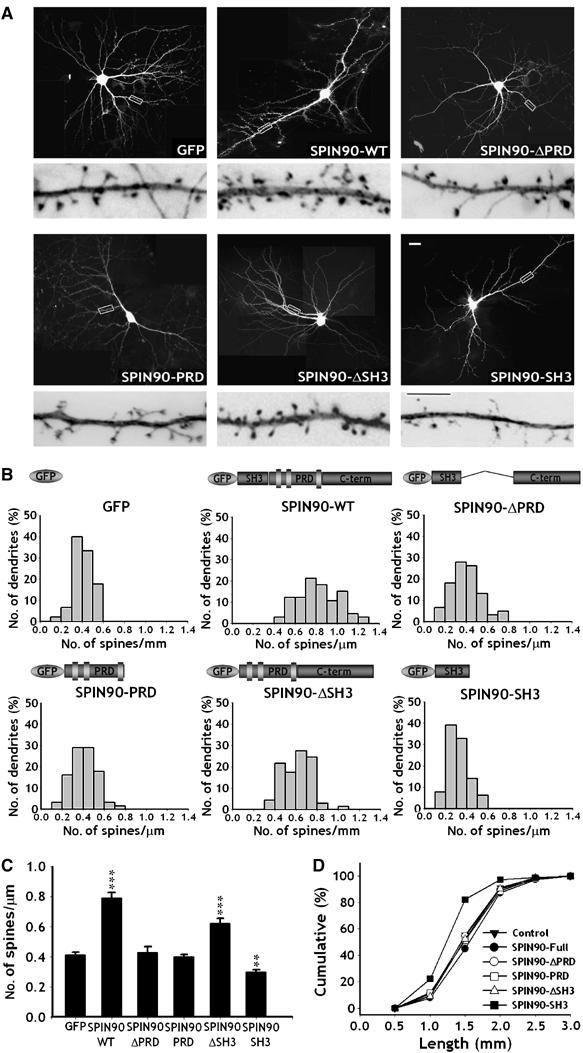
Overexpression of SPIN90/WISH increases the density and length of dendritic spines in mature hippocampal neurons. Neurons were transfected and imaged as in Figure 3. (A) GFP fluorescence images of the transfected neurons. High-magnification views are of the regions enclosed in rectangles. Images are inverted for clarity. Scale bars, low magnification: 20 μm; high magnification: 5 μm. (B, C) Histograms of the number of dendritic spines per μm in the transfected neurons (B); comparison of the average numbers of spines (C). Data are means±s.e. (GFP, n=6; SPIN90/WISH, n=8; SPIN90/WISH-ΔPRD, n=7; SPIN90/WISH-PRD, n=6; GFP-SPIN90/WISH-ΔSH3, n=7; GFP-SPIN90/WISH-SH3, n=7; ***P<0.01 and **P<0.05, significantly different from the GFP alone by ANOVA and Tukey's HSD post hoc test). SPIN90-ΔSH3, which lacks an N-WASP binding site, increased spine density like full-length of SPIN90/WISH, indicating an N-WASP-independent mechanism. (D) Cumulative frequency distribution of dendritic spine lengths in the transfected neurons.