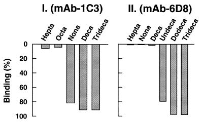
Figure 4.
Mapping of the mAb-6D8 and mAb-1C3 epitopes by using synthetic peptides. Individual peptides (Table 1) were incubated in the presence of antibody and protein-A Sepharose beads as in Methods. After centrifugation, the presence of peptides in the supernates was analyzed by HPLC. The binding of each peptide to either antibody was measured as the decrease in the peptide peak intensity relative to parallel controls in the absence of antibody. The extents of binding are shown in bars as percent of the control.

