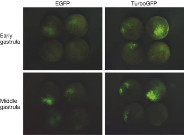
Figure 2.

Comparison of TurboGFP and enhanced green fluorescent protein maturation speed in developing Xenopus laevis embryos. At the stage of two blastomeres, embryos were microinjected with TurboGFP-C1 and pEGFP-C1 vectors. Living embryos were photographed from the animal pole side at the early and mid-gastrula stages. EGFP, enhanced green fluorescent protein.
