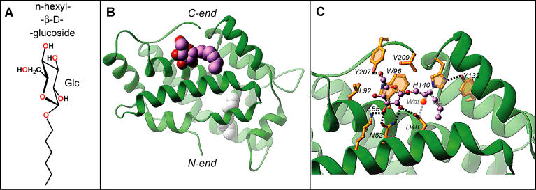
Figure 5. Structure of the n-hexyl-β-d-glucoside-GLTP Complex.
(A) Chemical formula of n-hexyl-β-d-glucoside.
(B) Crystal structure of the n-hexyl-β-d-glucoside-GLTP complex, with the n-hexyl-β-d-glucoside molecule accommodated within the sugar recognition center on the GLTP surface. The GLTP is shown in a green ribbon representation, and the carbon atoms of the n-hexyl-β-d-glucoside are shown in a lavender space-filling representation. Extraneous hydrocarbon is shown in a white space-filling representation.
(C) The n-hexyl-β-d-glucoside interactions with GLTP recognition center residues. Hydrogen bonds are shown by dashed lines. The bound ligand atoms are colored by lavender, red, and blue for carbon, oxygen, and nitrogen atoms, respectively. The water molecule bridging H140 with D48 is shown by bright red sphere.

