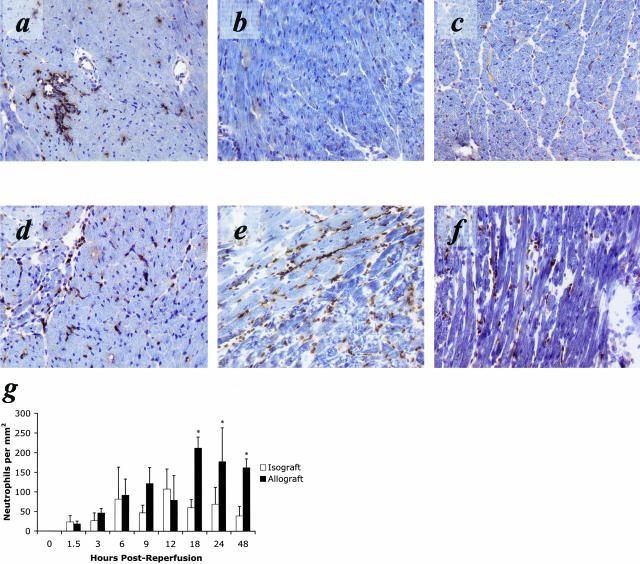
Figure 2.
PMN infiltration into cardiac allografts is increased in magnitude and duration compared to PMN infiltration observed in cardiac isografts controls. Immunohistology with anti-Ly6G (RB6–8C5) demonstrates an equivalent PMN infiltrate in cardiac isografts (a) and cardiac allografts (d) at 12 hours post-reperfusion. Sections taken at 24 hours post-reperfusion demonstrate the resolution of inflammation in isografts (b) but an enhanced PMN infiltrate into allografts (e). This PMN dominant inflammation is maintained at 48 hours in the allograft (f), while the isograft tissue (c) at this time appears normal. g: Quantification of immunohistology demonstrates that PMN infiltration into isografts (□) peaks around 12 hours post-reperfusion and returns to low levels by 24 hours post-reperfusion. However, PMN infiltration into allografts (▪) increases to twice that level between 12 hours and 24 hours post-reperfusion and remains elevated beyond 48 hours post-reperfusion (*, P < 0.01). All results are displayed as the mean and SD of at least 10 random myocardial fields from two non-consecutive sections from three to four grafts per group counted twice in a blinded fashion. The counts are normalized to tissue area and shown in mm2.

