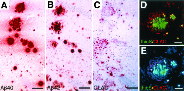
Figure 5.
Immunohistochemisty of the hilar region of dentate gyrus of the hippocampus in PSAPP TG mouse (19-month-old). Six-μm thick serial sections were immunostained for Aβ40 (BA27, A), Aβ42 (BC05, B), and CLAC (anti-NC2–2, C), and then counterstained with hematoxylin. Bar, 100 μm. D–E: Triple immunofluorescence labeling of cored plaques and surrounding diffuse deposits for thioS, CLAC, and Aβ. ThioS-positive core (green) and CLAC-positive deposits in the surrounding areas (red) are shown (D). E: Superimposition of immunoreactivity for human Aβ (blue, BAN50) shows that areas positive for CLAC (purple) or thioS (light green) are Aβ-positive. Bar, 50 μm.

