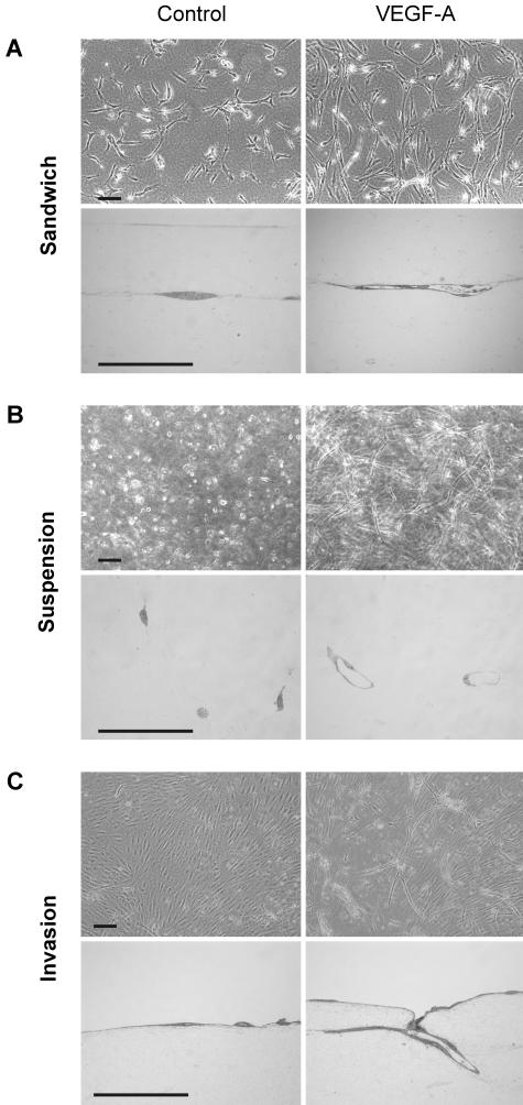
Figure 6.
Tube formation by hTERT-HDLEC and HMEC-1 in collagen gels. Cells were either sandwiched between two collagen gels (A), seeded in suspension as isolated single cells in a collagen gel (B), or seeded on the surface a collagen gel (C). Cells were left untreated (left) or treated with the indicated cytokines (right). After 5 to 7 days of culture, cells were photographed, and semi-thin sections (bottom) were analyzed to assess whether tube formation had occurred. Bar, 100 μm. Endothelial cell responses to cytokines are summarized in Table 2.

