Figure 5.
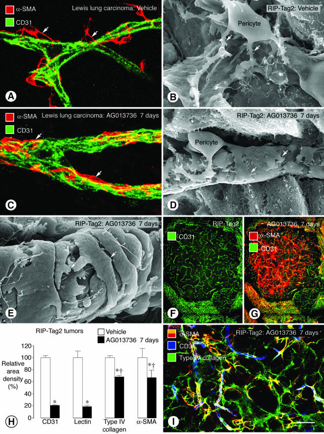
Tightening of pericytes on tumor vessels after inhibition of VEGF signaling. Confocal microscopic and SEM images show changes in pericyte-endothelial cell relationships (arrows) in tumor vessels. In vessels of LLC, α-SMA immunoreactive pericytes (A, red) are loosely attached to CD31-positive endothelial cells (green). A similar loose association of pericytes to endothelial cells is also evident by SEM in RIP-Tag2 tumor vessels (B). By comparison, after treatment with AG013736 for 7 days, α-SMA-positive pericytes are tighter on endothelial cells imaged by immunofluorescence (C) or SEM (D). Pericytes on some treated vessels acquire a smooth muscle-like phenotype (E). RIP-Tag2 tumor vessels still present after AG013736 for 7 days (F) are accompanied by a disproportionately large number of α-SMA-positive cells, many of which are not associated with blood vessels (G). After treatment with AG013736, reductions in α-SMA and type IV collagen immunoreactivities are about the same but are much less than corresponding reductions in CD31 and lectin (H, values for vehicle normalized to 100%). After the ∼80% reduction in RIP-Tag2 tumor vessels treated with AG013736 for 7 days, most α-SMA-positive cells are still covered by type IV collagen basement membrane (I). *, Different from corresponding vehicle (P < 0.05). †, Different from CD31. Scale bar: 20 μm (A, C); 5 μm (B, D); 500 μm (F, G); 50 μm (I).

