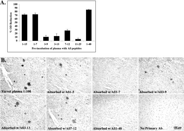
Figure 3.
Epitope-mapping of vervet anti-Aβ antibodies. A: A competitive ELISA was performed by pre-incubating immunized vervet plasma (16-year-old male at Day 301) individually with short Aβ peptide fragments. Bars represent the percent reduction in OD. Pre-incubation of plasma with Aβ peptides 1–15, 1–7, and 1–40 resulted in a 70% or greater reduction in OD, indicating that these peptides bound most of the Aβ antibodies in plasma and thus were not available to bind the plate-bound Aβ1–40. B: Sections from the frontal cortex of a 76-year-old female AD patient were immunostained with the same vervet plasma after pre-incubation with each of ten different Aβ peptides (Aβ1–5, 1–7, 1–15, 3–9, 3–13, 6–20, 7–12, 11–25, 26–42, and 1–40). Figure 3B shows representative examples of the results. Plaque staining was abolished by pre-incubation of vervet plasma with Aβ1–7, 1–15, or 1–40 peptides, indicating that the plasma antibodies bound the peptides during pre-incubation and were unavailable for binding to plaque Aβ protein. Plaque staining was observed with plasma diluted at 1:100 or absorbed with Aβ1–5, 3–9, 3–13, 6–20, 7–12, 11–25, 26–42. Plaque labeling was absent when primary antibody was omitted.

