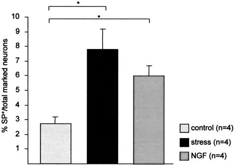
Figure 8.
Percentage of SP-positive neurons among the total number of marked neurons in the dorsal root ganglia taken from C4 to Th10 in non-stressed control mice, stressed mice, and non-stressed mice subcutaneously injected with NGF. On average, 14 ganglia were taken per mouse, and 40 consecutive slides were evaluated to obtain the present data, which are depicted as mean ± SEM. *, P < 0.05.

