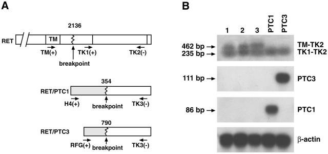
Figure 2.
A: A schematic of the primers selected for the RT-PCR. B: Detection of RET, RET/PTC1, and RET/PTC3 mRNA expression by RT-PCR. Lane 1, MTC tissue of patient 2; lane 2, PTC tissue of patient 2; lane 3, PTC tissue of patient 3: both transmembrane and tyrosine kinase domains are amplifiable, indicating the presence of full-length RET mRNA. Lanes PTC1 and PTC3, positive controls represented by cell lines expressing either one of the rearranged oncogenes: only the tyrosine kinase domain is amplifiable, indicating the presence of RET rearrangements.

