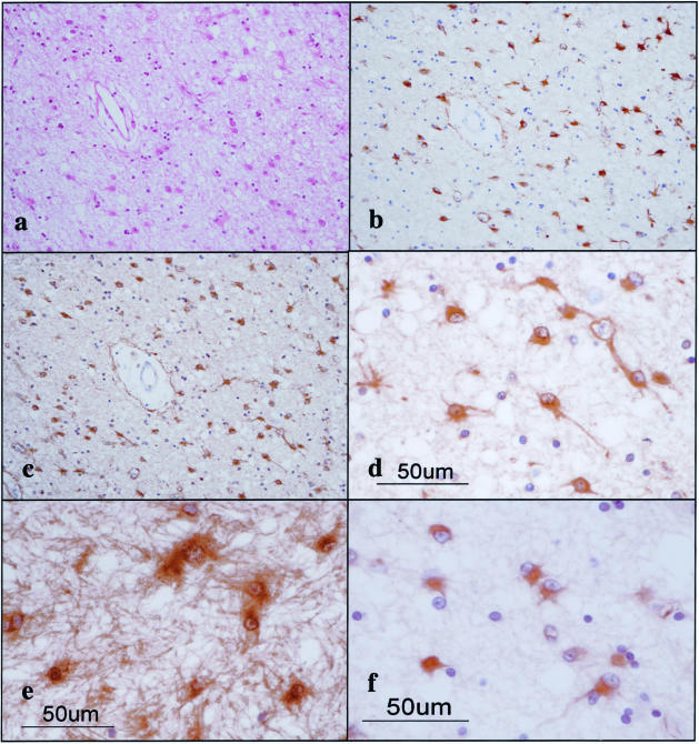
Figure 3.
The 14-3-3 ε isoform is expressed in reactive astrocytes in chronic demyelinating lesions of MS. MS brain tissues were processed for immunohistochemical analysis using ε isoform-specific antibody or the antibody against GFAP or vimentin. a to f represent the following: a: no. 744 MS, chronic active demyelinating lesions in the subcortical white matter of the frontal lobe (H&E). b: No. 744 MS, the area corresponding to a (GFAP). Many reactive astrocytes are stained. c: No. 744 MS, the area corresponding to a (ε). Many reactive astrocytes are stained. d: No. 744 MS, a higher magnification view of c (ε). Reactive astrocytes are stained. e: No. 544 MS, chronic inactive demyelinating lesions in the optic nerve (ε). Reactive astrocytes and the glial scar are stained. f: No. 744 MS, chronic active demyelinating lesions in the subcortical white matter of the frontal lobe (vimentin). Reactive astrocytes are stained.

